
 To the right is an untouched Mackerel fish. These fish when alive inhabit tropical and temperate waters, and are characterised by the vertical stripes on their backs, as well as their deeply forked tails.
These stripes surprisingly enough, are not used as camouflage, but rather as visual cues when these fish form schools with other members of the species. As their eyes are sensitive to moving stripes, these patterns allow the fish to follow each other and thus move efficiently in a group, as an anti-predatory adaptation.
 The first incision to the fish is seen here, being administered by my lab partner along the bottom of the body; originating at the anus. He goes on the run the scissors along the full length of the fish, before cutting a few centimetres higher along the top length of the fish. This allows the skin to be neatly peeled back, revealing the inside.
However before doing so, we first peeled back some of the skin covering the head and pharynx, and had a look at the gills that lie underneath (the red structures pictured). As fish take in water through the mouth, the liquid rushes across these filament-like structures which are covered in capillaries (tiny vein-like structures full of blood). These capillaries absorb the oxygen in this water and transfer it to the bloodstream, which gets pumped through the rest of the body.
 Carbon dioxide, which the fish must remove, is also passed through these capillaries. But rather than being pumped through the body, is transferred from the body to this same water entering the mouth. As the water gets expelled out of the gills and into the surrounding water, it takes this excess carbon dioxide with it. (Microscopic view of gill below)
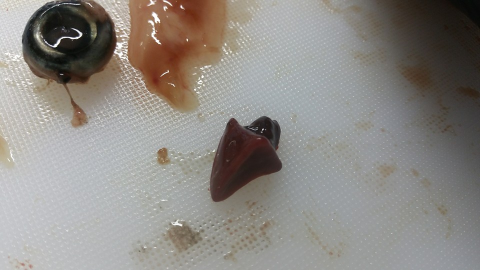 After further dissection, we found the fish heart (triangular red structure pictured), a muscular organ used of course to pump blood throughout the body. To its far left is whats left of the mackerel eye.
Zoology: sticky, slimy, and gross at times. But no one can ever say it isn't interesting (me in middle, along with high-spirited fellow students)

-Thomas Glen
Facebook.com/goodnaturepage
No comments:
Post a Comment